Microscopical
Society of Southern California
2004 Meeting Program
|
|
Microscopical
Society of Southern California
2004 Meeting Program
|
Weds January 21, 2004 at 7.30pm, New Roads School (map)
|
Weds February 18, 2004 at 7.30pm, New Roads School (map) This month's meeting will be a swap-meet or buy and sell meeting. Please bring any items you would like to offer for sale. There will also be a brief presentation on chemical crystal mounting and preparation techniques. |
|
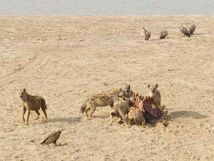
At this meeting, Larry Albright will give a presentation on his latest African photo Safari. He will present images of animals taken during the trip and will describe how one can acquire such images of animals using digital equipment. After this, Alan deHaas will give another talk in his lecture series on the technology of the microscope, this one focusing on binocular optical systems. |
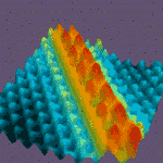
Weds April 21, 2004 at 7.30pm, New Roads School (map) At this meeting, Dr. Shijie Wu will present a program entitled "Scanning Probe Microscopy under Controlled Environments". Dr. Wu received his PhD. from the University of Guelph, Canada, in the area of physical chemistry, observing the structure of the solid-solution interface using in-situ x-ray diffraction, Atomic Force Microscope (AFM) imaging, and electrochemistry. He joined Molecular Imaging - a developer and supplier of AFM and Scanning Probe Microscope systems - as an applications scientist in 1998. His presentation will focus on Scanning Tunneling Microscope (STM) and AFM imaging under controlled conditions in the life science, material science, and nanotechnology fields, including some of Molecular Imaging's more recent developments, PicoPlus™, PicoTREC and MacMode.
Many polymer blends are very sensitive to environmental and temperature conditions. He will discuss experiments in which the humidity, the temperature, the solution composition or a combination of these parameters affects the topography and the physical properties of the surface. He will also present some new applications combining temperature and environmental control, as well as developments for local conductivity and impedance measurements used in battery and fuel cell studies. AFM is the only technique available today that can image, probe, and manipulate biological structures in environments simulating in-vivo conditions. Living cells can be imaged without fixation in buffers at 37°C. By placing an AFM on an inverted light microscopy, one can combine the information from light techniques with AFM measurements. Examples will be given in in-situ imaging of biological species ranging from cells to molecules, probing local properties such as elasticity, charge and conductivity, and manipulation of structures such as pulling or moving bio-molecules. |
Weds May 19, 2004 at 7.30pm, New Roads School (map)
|
At this meeting, Dr. Jack Green , Professor of Geological Sciences, Department of Geological Sciences, California State University, Long Beach will give a presentation entitled "A Semester of Optical Mineralogy in About an Hour." After a survey of light phenomena – Snell’s Laws, critical angles and Brewster’s Law, the details of the operation of the Nikon petrographic microscope are covered. A review of some thin sections introduces the student to basic vocabulary: analyzer and polarizer, mineral textures, relief, index of refraction, pleochroism, birefringence, retardation, etc. We then divide minerals into three groups: isotropic, uniaxial and biaxial with their respective 1, 2 and 3 indices of refraction stressing that the index of refraction is the reciprocal of the velocity of light in a specific direction in a given mineral. Calibrated immersion liquids (oils) are used to measure these indices using Beck line movement. Relief (how visible the mineral appears) is detailed. Diamond has a very high relief; fluorite very low. Another diagnostic feature is extinction where a mineral will extinguish (go black) with crossed “nicols” at specific angles as the stage is turned. Also under crossed nicols, interference colors can be seen due to subtraction of certain colors from white light. One of two rays that reachs the eye is faster or slower than the other. This is explained by the atomic structure of the mineral; calcite being an excellent example in explaining this fundamental observation. Accessory plates (gypsum, mica, quartz) inserted into the microscope light path enable the distinction of what is a fast or slow ray. The optical sign of a mineral is determined using (1) the Bertrand lens which creates a light pattern called an interference figure and (2) accessory plates. Details will be provided. There are relationships between mineral thickness, birefringence and retardation using a Michel-Levy color chart. Light in an anisotropic mineral can be modeled using an ellipsoid construct called an indicatrix whose principal axes represent indices of refraction. In uniaxial minerals there is one direction (coincident with a vibration axis) along which there is only one velocity; in biaxial minerals there are two such axes not coincident with major vibration axes. The angle between the optic axes in biaxial minerals is called 2 V and can be used to identify the mineral under certain conditions. Sometimes color fringes bordering isogyres can be seen in specific interference figures. In all of these optical procedures the student must always pat attention to mineral associations, cleavage, color and alteration. |
| Weds July 21, 2004 at 7.30pm, New Roads School (map)
|
Weds August 18, 2004 at 7.30pm, New Roads School (map)
The second half of the meeting will be a presentation (courtesy of Stuart Warter) of a new computer program called Micro-CT, Gateway to the 3-D Microworld. |
Weds September 15, 2004 at 7.30pm, New Roads School (map) This will be an exhibition event dedicated to photomicrography. Members are invited to show off their own images acquired through the microscope. Please be prepared to describe the image, as well as details of how it was obtained. There is no limit on how many images you may exhibit. |
Weds October 20, 2004 at 7.30pm, New Roads School (map)
|
Weds November 17, 2004 at 7.30pm, New Roads School (map) This is the annual Exhibition Meeting of the Society. This is one of the best events of the year and is a great deal of fun. Each member is encouraged to bring along an exhibit to share. Anything associated with microscopic subjects is welcome. Your exhibit could be simple, for example you could set up your microscope with your favorite slide. A projector will be provided for those bringing 35mm slides. Posters and display boards are also encouraged, along with the usual sales table. Please remember to bring a label or piece of paper with a brief description of your exhibit. |
No December meeting, instead the MSSC Holiday Banquet, December 15, 2004 at Earth, Wind and Fire, 2222 Wiltshire Blvd, Los Angeles. Reservations and prepayment required. |
WHAT'S
NEW? / MSSC HOME PAGE / MSSC
HISTORY / PROGRAM SCHEDULE /
ITEMS FOR SALE / NEWS
AND EVENTS / ARTICLES & RESOURCES / CONTACT US / HOW TO JOIN / LINKS / MEMBERS
AREA
Cartoons by Nirvan Mullick
Site created and maintained by Leonie
Fedel
Please email comments
© MSSC
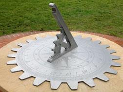 This
month MSSC member, Larry McDavid, will present a program entitled "SUNDIALS:
Prehistory to the Digital Age." In addition to being an MSSC member,
Larry is also a member of the North American Sundial Society, which -
along with the British Sundial Society - actively promotes the study,
development and construction of these light-operated calendar and time
instruments. Larry will discuss the history and science of dialing and
show pictures of many kinds of sundials throughout the world. If you have
an interest in sundials, we invite you to bring along your dials or other
solar instruments for display and discussion. There will be a table set
up for the purpose of exhibiting the instruments. Larry's talk will be
supported with a PowerPoint presentation of 120 slides illustrating the
subject. Most of the images will be presented in a section called "Beauty
in Dialing" and will run as a continuous slide show during the break.
This
month MSSC member, Larry McDavid, will present a program entitled "SUNDIALS:
Prehistory to the Digital Age." In addition to being an MSSC member,
Larry is also a member of the North American Sundial Society, which -
along with the British Sundial Society - actively promotes the study,
development and construction of these light-operated calendar and time
instruments. Larry will discuss the history and science of dialing and
show pictures of many kinds of sundials throughout the world. If you have
an interest in sundials, we invite you to bring along your dials or other
solar instruments for display and discussion. There will be a table set
up for the purpose of exhibiting the instruments. Larry's talk will be
supported with a PowerPoint presentation of 120 slides illustrating the
subject. Most of the images will be presented in a section called "Beauty
in Dialing" and will run as a continuous slide show during the break.
 Dr.
Wu will discuss how many experiments, particularly biological ones, benefit
from imaging under controlled conditions. Here, electrochemical measurements
are carried out in solution, mostly with the absence of dissolved oxygen.
He will explain, how by combining electrochemical control with scanning
probe microscope, one can manipulate an electrode surface and study the
changes at real time with resolutions ranging from atomic to micron scales.
Dr.
Wu will discuss how many experiments, particularly biological ones, benefit
from imaging under controlled conditions. Here, electrochemical measurements
are carried out in solution, mostly with the absence of dissolved oxygen.
He will explain, how by combining electrochemical control with scanning
probe microscope, one can manipulate an electrode surface and study the
changes at real time with resolutions ranging from atomic to micron scales.
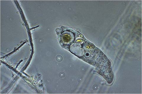 The
Pond Life program is undoubtedly one of our best attended and most exciting
meetings! Members are strongly encouraged to bring pond water, ditch water,
fountain water or anything that lives in water. Microscopes, illuminators
and pipettes and tools to play in the water are required.(Extra microscopes
are always a good thing to bring for guests to use.) Check out the amoeba
to the right, courtesy of
The
Pond Life program is undoubtedly one of our best attended and most exciting
meetings! Members are strongly encouraged to bring pond water, ditch water,
fountain water or anything that lives in water. Microscopes, illuminators
and pipettes and tools to play in the water are required.(Extra microscopes
are always a good thing to bring for guests to use.) Check out the amoeba
to the right, courtesy of  Weds
June 16, 2004 at 7.30pm, New Roads School (
Weds
June 16, 2004 at 7.30pm, New Roads School ( At
this meeting, Dr. Rick Behl, Associate Professor of Geological Sciences,
from California State University, Long Beach, will give a presentation
entitled, "Analysis of sea bed cores and implications for climate
change." Sediments are deposited and therefore help us understand
the surface of the Earth. If you want to understand how the Earth's climate,
oceans and geography have changed over time, the sedimentary record is
the only place that holds a fuller perspective than the short historical
record of humans. Sedimentologists learn to read the structures and textures
of sedimentary rocks to uncover their ancient secrets. Dr. Behl's research
focuses on the sedimentology of modern and ancient continental margins
and deep-sea upwelling systems, which are particularly good recorders
of ancient environments. The organic-rich sediments deposited in these
places play a key role in the global carbon cycle, climatic regulation,
and the formation of oil and gas. His recent research deals with abrupt
environmental shifts along the California margin linked to climatic and
tectonic change.
At
this meeting, Dr. Rick Behl, Associate Professor of Geological Sciences,
from California State University, Long Beach, will give a presentation
entitled, "Analysis of sea bed cores and implications for climate
change." Sediments are deposited and therefore help us understand
the surface of the Earth. If you want to understand how the Earth's climate,
oceans and geography have changed over time, the sedimentary record is
the only place that holds a fuller perspective than the short historical
record of humans. Sedimentologists learn to read the structures and textures
of sedimentary rocks to uncover their ancient secrets. Dr. Behl's research
focuses on the sedimentology of modern and ancient continental margins
and deep-sea upwelling systems, which are particularly good recorders
of ancient environments. The organic-rich sediments deposited in these
places play a key role in the global carbon cycle, climatic regulation,
and the formation of oil and gas. His recent research deals with abrupt
environmental shifts along the California margin linked to climatic and
tectonic change.  At
this meeting, Gregg Kleinberg returns to give a talk on digital photomicrography.
Mr. Kleinberg has a great deal of experience in this field. Over the years
he has come to the conclusion that the
At
this meeting, Gregg Kleinberg returns to give a talk on digital photomicrography.
Mr. Kleinberg has a great deal of experience in this field. Over the years
he has come to the conclusion that the 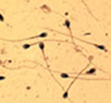 At
this meeting, MSSC member, Edwin Jones, will give a presentation entitled,
"The Morphological Identification of Sperm Heads: Two Death Penalties,
A Civil Suit and a Chapter." This talk will take the audience through
experiences that defend the use of sperm heads to identify semen. In 1998,
Orange County required verification of some sperm heads found as evidence
in a homicide involving a body that had been kept in a freezer by the
suspect for three years. The case against the suspect was very strong;
he was caught with the victim's body and many of her personal possessions.
Her blood was located at the suspect's place of business. The Orange County
Sheriffs Crime Lab had identified sperm heads in anal samples from the
victim, but only the victim's DNA was found in this sample. The defendant
admitted the homicide but denied having sex with the victim. The defense
brought in the retired director of an out-of-state crime lab and an Ivy
League fertility doctor who testified that an intact sperm (head attached
to tail) is needed for identification. Mr. Jones testified that the sperm
heads were identifiable without an attached tail. To find out the jury's
verdict in this case, and two other cases involving the identification
of sperm heads, in which Mr Jones was asked to testify as an expert witness,
come along to this month's Society meeting.
At
this meeting, MSSC member, Edwin Jones, will give a presentation entitled,
"The Morphological Identification of Sperm Heads: Two Death Penalties,
A Civil Suit and a Chapter." This talk will take the audience through
experiences that defend the use of sperm heads to identify semen. In 1998,
Orange County required verification of some sperm heads found as evidence
in a homicide involving a body that had been kept in a freezer by the
suspect for three years. The case against the suspect was very strong;
he was caught with the victim's body and many of her personal possessions.
Her blood was located at the suspect's place of business. The Orange County
Sheriffs Crime Lab had identified sperm heads in anal samples from the
victim, but only the victim's DNA was found in this sample. The defendant
admitted the homicide but denied having sex with the victim. The defense
brought in the retired director of an out-of-state crime lab and an Ivy
League fertility doctor who testified that an intact sperm (head attached
to tail) is needed for identification. Mr. Jones testified that the sperm
heads were identifiable without an attached tail. To find out the jury's
verdict in this case, and two other cases involving the identification
of sperm heads, in which Mr Jones was asked to testify as an expert witness,
come along to this month's Society meeting.