Microscopical
Society of Southern California
2010 Meeting Program
|
|
Microscopical
Society of Southern California
2010 Meeting Program
|
Weds January 20, 2010 at 7:00pm, New Roads School (map) MEETING CANCELLED DUE TO WINTER STORM. Instead members are invited on a field trip, January 30, 2010 to Ventura County Sheriff's Department Forensic Crime Lab. See members area for further information. |
||
Weds February 17, 2010 at 7:00pm, New Roads School (map)
|
||
| Weds
March 17, 2010 at 7:00pm, New Roads School (map) For the second in our mini series on specialized
contrast techniques for the light microscope, at this meeting Dr. Brian Matsumoto (UCSB) discuss Confocal Microscopy. In standard microscopes, light arising from outside the plane of focus will be projected
on the imaging sensor--be it the human eye or a digital detector. For work with
transmitted illumination, this is of little import and a high contrast view of a transparent
specimen can be obtained. In contrast, for thick fluorescent material, out-of-focus light
will obscure the view by generating glare. Confocal microscopes remove this with an
aperture sized and positioned to pass only light arising from the plane of focus.
This high contrast view is an optical section. Dr. Matsumoto will present micrographs obtained
with this technology and show their advantages in biological and three- |
||
Weds April 21, 2010 at 7:00pm, New Roads School (map)
|
||
Weds
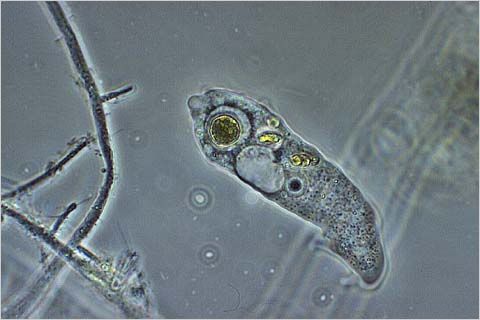
May 19, 2010 at 7:00pm, New Roads School (map) The Pond Life program is undoubtedly one of our best attended and most exciting meetings! Members are strongly encouraged to bring pond water, ditch water, fountain water or anything that lives in water. Microscopes, illuminators and pipettes and tools to play in the water are required.(Extra microscopes are always a good thing to bring for guests to use.) Check out the amoeba to the right, courtesy of www.micrographia.com. |
||
Weds June 16, 2010 at 7:00pm, New Roads School (map)
The opening presentation will review a subject that can very often greatly affect our personal comfort - allergens. Depending on your level of tolerance for allergens, you may find it fascinating that this year (2010) has been the worst year in recorded history for massive pollen counts. Mother Nature has responded in a big way to the rather late arrival of spring. We will look at how pollen is counted and just where it comes from and what it looks like. This is a major public service effort that still very much depends on the use of the microscope. |
||
| Weds July 21, 2010 at 7:00pm, New Roads School (map)
Digital SLRs have been used as photomicrography cameras and while they
have the virtue of being inexpensive and readily available, scientists have
preferred working with dedicated cameras designed specifically for
photomicrography. Although such cameras tend to be more expensive,
their advantages justify their cost and for the serious user, they are the
preferred instrument. Especially for work that requires generating large
numbers of images, the dedicated cameras, such as the PAXcams, are
invaluable because they provide an ergonomic and convenient tool for the
microscopist. Such cameras do not have a mechanical shutter and they
provide a vibration free operation that ensures maximum sharpness.
Moreover, they communicate directly with a computer and provide an
enlarged image of the specimen. Precision focusing is much more easily
attained with this arrangement and it is easier and more convenient to achieve
high quality photomicrographs with such a camera. For quantitative work
with image processing and for quantitative analysis such cameras are the
preferred tools. To mount a dedicated digital cameras the majority of research
microscopes are equipped with "c" mounts. These are an industrial standard for
electronic cameras and consist of a male screw with a 25 mm bore. This limits
the pixel array of a camera and many of these are 2 mega-pixels in size.
This is not a disadvantage for scientific work; however, it can be a limitation
for photographers who create images for magazine reproductions. |
||
We're turning this meeting in a Swap Meet. We are hoping to have all the tables in the room full of items available for sale. This is your chance to sell things that you no longer need and the chance to find items that you may have been looking for. |
||
Weds
September 15, 2010 at 7:00pm, New Roads School (map) This week Damara Gebauer, Field Application Scientist from Cambridge Research Instrument Division will discuss some of the newest technology in multispectral imaging in microscopy. A very new and advanced system has been developed for extending the advantages of fluorescence (and other) methods of using image markers. This new technology almost completely eliminates the problems of signal to noise in resolving labeled specimens. CRI’s multispectral imaging systems enable users to quantitate molecular markers even when they are co-localized in a single tissue section, producing clear and accurate images of each individual label on a multi-label tissue section. These systems also offer the powerful capability to unmix and remove autofluorescence in fluorescence images, thereby dramatically increasing signal-to-noise and improving the accuracy of your results. Nuance systems deliver flow-cytometry-like data while preserving the morphological context down to the sub-cellular level.” InForm™ is a train-by-example software platform that can be readily trained to separate image regions into appropriate classes (‘cancer’, ‘stroma’, ‘inflammation’, e.g.) with unprecedented accuracy. It can be combined with specific segmentation and quantitation tools to extract molecular data automatically from appropriate cellular and tissue compartments, information necessary for designing and testing targeted diagnostic and therapeutic reagents. |
||
Weds October 20, 2010 at 7:00pm, New Roads School (map) MSSC member, Stuart Warter - an expert in the art of nature photography - will kick off this meeting with a presentation of some of his photographs of birds. For the rest of the evening, we plan on something a little different, something that will require the actual use of a microscope. Allan has acquired a marvelous collection of slides illustrating parasites. Alan will show the slides on the microscope using a video camera and monitor. There will be a discussion on the contents of the mounts. |
||
Weds November 17, 2010 at 7:00pm, New Roads School (map) This is the annual Exhibition Meeting of the Society. This is one of the best events of the year and is a great deal of fun. Each member is encouraged to bring along an exhibit to share. Anything associated with microscopic subjects is welcome. Your exhibit could be simple, for example you could set up your microscope with your favorite slide. A projector will be provided for those bringing 35mm slides. Posters and display boards are also encouraged, along with the usual sales table. Please remember to bring a label or piece of paper with a brief description of your exhibit. Also note that on November 6, 2010 the Hammer Museum in Los Angeles wil be hosting the Enormous Microscopic Evening, an interactive exhibit of Machine Project This free public event dedicated to performed microscopy will feature demonstrations of research and equipment from scientists, technical innovators and amateurs who use these instruments as an integral aspect of their work. Participants include representatives from Stanford's Clark Center, the Fletcher Lab at UC Berkeley, the LA Museum of Natural History, USC and the San Francisco Exploratorium amongst others. In addition to demonstrations and activities that will bring attention to the field of microscopy this program will have screenings and a workshop on how to build a web cam microscope and the original Leeuwenhoek viewing apparatus. |
||
No December meeting, instead the MSSC Holiday Banquet, December 12, 2010, 5:00 - 9:00 p.m. The Holiday Banquet is set for Sunday, December 12th 2010. Come and celebrate the completion of another wonderful year with fellow MSSC members. The gathering will be at the Earth Wind & Flour Restaurant, 2222 Wilshire Blvd., Santa Monica, CA 90403. Phone 310 829-7829. Please bring cash as there will be no individual checks. Dinners are about 10-20 dollars depending on what you order from the menu. The program will include our annual report and by request a colorful Christmas show represented by images never yet seen taken by the master of photomicrography, John Chesluk. Please rsvp to MSSC President, Jim Solliday if you plan on attending. jlsolliday @ roadrunner.com. |
WHAT'S
NEW? / MSSC HOME PAGE / MSSC HISTORY / PROGRAM SCHEDULE /
ITEMS FOR SALE / NEWS
AND EVENTS / VIDEOS, ARTICLES & RESOURCES / CONTACT US / HOW TO JOIN / LINKS / MEMBERS
AREA

Cartoons by Nirvan Mullick
Site created and maintained by Leonie
Fedel
Please email comments
© MSSC


 Saturday,
August 18, 2010 at 7:00pm, New Roads School (
Saturday,
August 18, 2010 at 7:00pm, New Roads School (