Microscopical
Society of Southern California
2012 Meeting Program
|
|
Microscopical
Society of Southern California
2012 Meeting Program
|
Note: ideas expressed by speakers at these meetings are their own, and do not necessarily represent those of the MSSC.
Our first gathering for 2012 will be a special meeting with guest speaker, Brian Ford, joining us from England. Brian is an authority on the work and history of A. van Leeuwenhoebe and will be giving a presentation on his latest study of van Leeuwenhoebe. The presentation will include some never-before-seen video sequences of exactly what Leeuwenhoek saw through his instruments. Don't miss this historic event. Brian is a world-class speaker who always provides entertaining and informative presentations. Please note: this meeting on the first TUESDAY of January will replace our normal third Wednesday gathering and will be our only lectureship event for the month of January. |
Weds February 15, 2012 at 7:00pm, New Roads School (map) MSSC resident expert on all things photographic, Brian Matsumoto (UCSB) and his wife Carol will bring us up to date on the latest digital imaging technology. Brian has been researching Sony camera technology and will share his findings, including which is the best SLR type camera for use with a microscope. Illustrations and examples will be provided. In the second half of the meeting MSSC President, Jim Solliday, will provide a "preview" of the work he is doing on the petrology of meteorites. He will show what the inside of meteorites look like through a polarizing microscope. Meteorites come in three types: iron, stony-iron and stony.The stony meteorites are quite splendid under polarized light. |
|
This meeting's guest speaker will be John Izbicki, presenting on "Sources of Fecal Indicator Bacteria along the Malibu, California, Coastline." Fecal indicator bacteria (FIB) are occasionally present at concentrations that exceed recreational water-quality standards in Malibu Lagoon and at ocean beaches near Malibu, California. Septic tanks are used to treat residential and commercial sewage in the area; consequently, discharge of shallow groundwater containing septic-tank effluent to the lagoon and beaches is a possible source of FIB. Radon-222, an indicator of groundwater discharge, measured during the dry summer season from July 21-27, 2009 showed discharge to the lagoon during high tide and discharge to the near-shore ocean during low tide. Samples collected from the lagoon during that period had total coliform, Escherichia coli, and enterococcus concentrations as high as 650,000, 130,000, and 3,400 MPN per 100 mL, respectively. However, human-specific Bacteroidales, an indicator of human-fecal contamination, were not detected in any of the samples. The absence of Bacteroidales in the lagoon and low FIB concentrations in shallow groundwater downgradient from septic tanks suggests that septic-tank effluent was not the source of high FIB concentrations in the lagoon during the sample period. Although the source of FIB in the lagoon was not identified, water movement from the lagoon through the sand berm at the mouth of the lagoon was indentified as a source of FIB to the adjacent near-shore ocean at low tide. The absence of Bacteroidales and low FIB concentrations adjacent to unsewered residential development at low tide suggests that groundwater discharge was not a source of FIB to the near-shore ocean at beaches not influenced by discharge from the lagoon |
This month we will have a meeting dedicated to trade and sales. Table space will be available to all present to share items for sale or trade. Members will have the opportunity to find something they have been looking for or need for a project. If you have stuff you wish to sell, bring it to the meeting, if you have equipment or supplies you are looking for then do come to the meeting and take a look around. Bring a little cash or your check book just in case you find that item you have been looking for. We would also like to encourage members to bring in items that are NOT for sale. For example, bring along that special item or exhibit, so members who have not been able to attend the Saturday workshops can see the best of the best of our Saturday displays. We will dedicate the last hour of our sales meeting to the exhibits and give workshop members the chance to describe the beautiful and rare items we enjoy on the first Saturday events. |
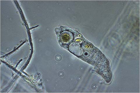
The Pond Life program is undoubtedly one of our best attended and most exciting meetings! Members are strongly encouraged to bring pond water, ditch water, fountain water or anything that lives in water. Microscopes, illuminators and pipettes and tools to play in the water are required.(Extra microscopes are always a good thing to bring for guests to use.) Check out the amoeba to the right, courtesy of www.micrographia.com. |
Weds June 20, 2012 at 7:00pm, New Roads School (map)
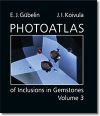
To fully identify an inclusion in a gem or mineral, including the complete analysis of its major and trace element chemistry, and a description of its structure, is a very time-consuming process. This type of inclusion work requires not only a solid understanding of gemology, but also a good foundation in chemistry and mineralogy. It also requires access to specialized equipment to do analytical procedures such as X-ray diffraction and quantitative chemical analysis.In order to appreciate this highly specialized skill, one must first know how inclusions are identified, and also what the inclusionist actually faces in doing this type of work. First and foremost, inclusion identification demands a considerable allotment of time. A high degree of specialized knowledge gained only through years of studying inclusions and trying to decipher their nature and meaning is also required for this type of work. Inclusion size is generally always a problematic fact that creates its own set of difficulties. It stands to reason that most inclusions we encounter are less than one half millimeter in size. While we do occasionally encounter incredibly large inclusions these are extreme rarities, and inclusionists are most often dealing with minute quantities of material, and working on a microscopic level. In this microworld the techniques used for inclusion work differ considerable from many of those taught in geoscience classrooms and laboratories. By their very definition inclusions are also difficult to work on because they are enclosed in their hosts, and protected by them. Completely sealed in, the overlying mineral shields inclusions from the inclusion analyst. The fact that gems and many minerals are valuable also limits what can be done to identify an inclusion. Generally speaking, any form of destructive analysis is most often out of the question. Another consideration that must be kept in mind is that a mineral or fluid inclusion might be exceedingly rare, unusual, or beautiful which would give the host an additional scientific or visual value that would add to its monetary worth. If such an inclusion is destroyed in order to identify it, then the visual value is lost. An inclusionist must therefore decide prior to analysis if the potential for loss is worth the potential scientific gain. Under the above considerations an inclusionist proceeds to attempt the identification of an inclusion as completely as time and the situation itself will allow. It happens on occasion that an inclusion is exposed to the surface by accidental breakage, or during the cutting process. Such instances prove fortunate for the inclusionist because this makes it easy to get at the inclusion, and because such gems and inerals are usually much less expensive that those which are not in this condition. However, it is more often the case that inclusions must be exposed to the surface or brought nearer to the surface of their host by some mechanical means in order to prepare them for identification. The methods of inclusion exposure most commonly employed include grinding and polishing, cracking, and crushing.“Gems” with interesting inclusions suitable for semi-destructive or destructive analysis, are not that easily obtained. This is because most of us do not have access to the large quantities of rough and cut gems, such as rubies, sapphires, emeralds, or diamonds that might yield useful analytical samples. Gems over 0.25 ct in weight with colorful or unusual inclusions would not be the primary targets for this type of research because of their potential value and visual appeal. Therefore most inclusion work of a destructive or potentially destructive nature is carried out on personally owned gems of very small size and often in poor condition. Gem and mineral collectors today value gems and transparent mineral crystals that contain brightly colored and interesting inclusions such as deep red cinnabar crystals in quartz or green diopside crystals in diamond. Those doing inclusion research usually cannot out bid or out buy “collectors” for gemstones or crystals with interesting inclusions. As a result, many such stones end up in private collections, and their inclusions, one of the reasons they were purchased in the first place, are never adequately documented. The “gem” materials most often used for inclusion research are generally those that have been rejected by the jewelry trade. Inclusion research is expensive, because gems are expensive, and because “Time is money.” Each gem sacrificed in the name of science represents at least a small financial loss to the owner, as well as a considerable expenditure of time, which is balanced only by the knowledge gained. When a gem is sacrifice for its inclusions there is really no guarantee that nything new or interesting will be learned. Such is the nature of inclusion research - as the saying goes, “When you explore the unknown, you know not what you will find.” Utilizing modern microscopic techniques and current technologies this lecture explores the hard-to-reach world of inclusions by introducing a number of new inclusion discoveries and observations in a wide variety of minerals and gems. For an introduction to John's work, see: http://www.microworldofgems.com/ --- During the second half of the meeting we will turn our attention to some photographs of the Transit of Venus that occurred on the 5th of June. In addition to showing some of the images captured by NASA, we will also show images taken by MSSC members including a splendid image by Brian Matsumoto, a professor from UCSB. MSSC member, Larry McDavid will also briefly describe the type of filters needed to study the sun. |
| Weds July 18, 2012 at 7:00pm, New Roads School (map) This month our guest speaker will be MSSC member, Brian Matsumoto (UCSB) who will give a talk on "Variations of Phase Contrast."
Phase contrast is a major advance in light microscopy and its development earned Frits Zernike the Nobel prize in physics(1953). Unlike other optical enhancing techniques, such as differential interference contrast, it is an affordable research tool that can be added to virtually any microscope with an interchangeable condenser. What is needed is a condenser that has a annular ring for generating a hollow cone of light and a modified objective lens that has a phase plate. When the plate and the phase ring are aligned, the microscope renders cells which are virtually featureless in brightfield illumination as high contrast images at high resolution. In spite of these advantages, the Heine apparatus was discontinued and Leitz adopted a Zernicke-type system. Instead of having an infinitely adjustable cone of light, they used a condenser with a set of fixed diameter phase rings. Like the Zeiss phase contrast system, the rings could be rotated into the optical path and their diameter was adjusted for a given numerical aperture and magnification. Leitz designed and manufactured a set of objectives (Phaco) for this condenser. We will provide images demonstrating the different quality images obtained by the Leitz and Zeiss microscopes. We will show that increasing the diameter of the phase rings can increase resolution of phase objects. This presentation will be filled with splendid images using a large variety of Phase systems manufactured by the top makers in Europe, don't miss this one. |
Weds, August 15, 2012 at 7:00pm, New Roads School (map) This month we will have the privilege of hearing from one of the areas most talented photomicrographers, David Scharf. His talk will be on The Art of SEM Photography, describing his imaging and specimen preparation techniques. Mr. Scharf has developed wonderful special affects in conjunction with the Scanning Electron Microscope, including colorization and 3D imaging. If you follow his work you will already know how impressive his images indeed are. His presentation will include both a computerized slide show and an exhibition of splendid prints. For a wonderful preview you should go to his website at the following: DAVID SCHARF PHOTOGRAPHY, Scanning Electron Microscopy http://www.scharfphoto.com or http://www.electronmicro.com. |
Birdwatchers have a hidden debt to history: the scientific study of birds took a specific route in the late nineteenth and early twentieth century that dictated the ways birds were understood and enjoyed. This meeting will feature a presentation by Daniel Lewis, the Huntington Library's Dibner Senior Curator of the History of Science and Technology, who will discuss his new book The Feathery Tribe, a biography of Robert Ridgway, the Smithsonian's first Curator of Birds. The book traces key changes in ornithology leading to the present - and what it meant to be a natural scientist at the dawn of the twentieth century. |
Weds October 17, 2012 at 7:00pm, New Roads School (map) This month, our speaker will be MSSC member, Brian Matsumoto (Prof. of Imaging Microscopy UCSB, Em). Our resident expert on imaging and camera technology has recently been asked to More color then a Christmas Tree: In this case we are actually talking about a tree, plant tissue under the microscope. Wood sections are made up primarily of cellulose and react positively when studied using interference filters. This not only reveals the morphology but renders every color in the spectrum visible to the eye. What makes this even more interesting is the preparations used are well over 100 years old. Many of them have technically gone bad and the question is are slides that have gone south still worth study? I think you will be amazed at the splendor and aesthetic appeal even bad mounts offer. Be ready for a kaleidoscope of colors and shapes. The title of this segment of our meeting will be the Antique Wood Sections of C.B. Johnson. |
Weds November 14, 2012 at 7:00pm, New Roads School (map) *** NOTE this is a week earlier than usual *** This is the annual Exhibition Meeting of the Society. This is one of the best events of the year and is a great deal of fun. Each member is encouraged to bring along an exhibit to share. Anything associated with microscopic subjects is welcome. Your exhibit could be simple, for example you could set up your microscope with your favorite slide. A projector will be provided for those bringing 35mm slides. Posters and display boards are also encouraged, along with the usual sales table. Please remember to bring a label or piece of paper with a brief description of your exhibit. |
No December meeting, instead the MSSC Holiday Banquet, date December 16, 2012 at Earth Wind and Flour 5 p.m. to 9 p.m. The Holiday Banquet is set for Sunday, December 16th 2012. Come and celebrate the completion of another wonderful year with fellow MSSC members. The gathering will be at the Earth Wind & Flour Restaurant, 2222 Wilshire Blvd., Santa Monica, CA 90403. Phone 310 829-7829. Please bring cash as there will be no individual checks. Dinners are about 10-20 dollars + 20% tip depending on what you order from the menu. RSVPs required by Dec. 7th to MSSC President, Jim Solliday jlsolliday @ roadrunner.com. |
Note: ideas expressed by speakers at these meetings are their own, and do not necessarily represent those of the MSSC.
WHAT'S
NEW? / MSSC HOME PAGE / MSSC HISTORY / PROGRAM SCHEDULE /
ITEMS FOR SALE / NEWS
AND EVENTS / VIDEOS, ARTICLES & RESOURCES / CONTACT US / HOW TO JOIN / LINKS / MEMBERS
AREA

Cartoons by Nirvan Mullick
Site created and maintained by Leonie
Fedel
Please email comments
© MSSC